Free guide
Free
The Midlife Essentials and the talk
A 75-page PDF guide plus the 1 hour 43 minute talk, The Entire Midlife Journey.
Send me the guide
By Daniela Hess, MSEd13 min readOctober 2026
Many Women get a screening letter in the mail and never learn what their breast tissue is made of. Here is what density means, why it can hide cancer on a mammogram, and what the 2024 FDA rule requires. You will also see what the trials show for ultrasound and MRI, and why every screening decision belongs to you and your practitioner.
Your breasts change in Midlife, and so do the questions about how to look after them.
Many Women get a screening letter in the mail and never learn what their breast tissue is made of or which other kinds of imaging exist. Breast care is more than 1 appointment a year or every 2 years. It includes knowing your own density, knowing your options, and tending your breasts at home.
This chapter covers breast density and the letter every Woman in the United States now receives about it, the full menu of imaging, from mammography to ultrasound, MRI, QT imaging, and thermography, what the evidence says about each, and the choices I have made for my own body. It ends with castor oil packs, the way I use them, and the questions to bring to your practitioner.
Your breasts follow the same arc as the rest of you. Alcohol in your 20's and 30's adds to breast cancer risk over the decades, and the body fat you carry into Menopause becomes a main source of the estrogen your breast tissue lives with afterward, and the screening and care you choose in your 40's and 50's shape what you live with at 60 and beyond.
In its 2025 advisory, the US Surgeon General estimated that about 16.4 percent of breast cancers in US Women in 2019 were linked to alcohol. Alcohol raises the estrogen your breast tissue lives with, and 'Why Alcohol No Longer Serves You', in 'Eating for Midlife: Protein, Fiber, Healthy Fats, Hydration, and Why Alcohol No Longer Serves You', explains how, and how to step away from it.
The Hormone Therapy 101 chapter already covers what the research shows about hormone therapy and breast cancer, so this chapter points back to it rather than repeating it.
This is education. Every screening decision is yours to make with your practitioner, with the facts in front of you.
A breast holds 2 main kinds of tissue. There is fibroglandular tissue: the milk glands, the ducts, and the fibrous connective tissue that holds them. And there is fat.
'Dense' breasts have relatively more glandular and fibrous tissue and relatively less fat. The National Cancer Institute is clear on 1 point that surprises many Women: you cannot feel density.
A dense breast does not feel firmer or lumpier. Only a radiologist looking at a mammogram can tell you whether your breasts are dense.
Radiologists sort breasts into 4 categories. These are the words the FDA now requires on your mammography report:
Categories C and D count as dense. Roughly 10 percent of Women fall into A, 40 percent into B, 40 percent into C, and 10 percent into D.
In a study of about 1.5 million mammograms, 43.3 percent of US Women aged 40 to 74 had heterogeneously or extremely dense breasts. Nearly half of the Women reading this book have dense breasts, and many do not know it.
Density shifts across a lifetime. According to the National Cancer Institute, breasts tend to become less dense with age and after having children, and Women who use menopausal hormone therapy or who have a lower body weight tend to have denser breasts. If you started hormone therapy since your last mammogram, your category may change, so let the imaging center know.
For years, whether a Woman was told about her density depended on which state she lived in. That changed on September 10, 2024, when an updated FDA rule under the Mammography Quality Standards Act took effect nationwide.
Now every mammography facility in the United States must tell you in writing whether your breast tissue is dense, and the report sent to your provider must name your density category.
If your breasts are dense, the FDA requires the letter to say this, word for word:
Breast tissue can be either dense or not dense. Dense tissue makes it harder to find breast cancer on a mammogram and also raises the risk of developing breast cancer. Your breast tissue is dense. In some people with dense tissue, other imaging tests in addition to a mammogram may help find cancers. Talk to your healthcare provider about breast density, risks for breast cancer, and your individual situation.
The federal government now tells Women with dense breasts, in writing, that other imaging tests may help find cancers.
The letter informs you. It does not schedule the next test, and it does not say which test. In a busy system, the follow-up conversation often waits for the Woman to start it.
Density matters for 2 separate reasons.
First, density can hide cancer. On a mammogram, fat looks dark, and dense tissue looks white.
A cancer also looks white. So a tumor in a dense breast can be like a snowball in a snowstorm.
The numbers show this masking. In data from US screening programs, the sensitivity of mammography, meaning the share of cancers it catches, fell from 81.2 percent in Women 40 to 49 with non-dense breasts to 63.3 percent in Women the same age with dense breasts. For Women 70 to 74, it fell from 90.0 percent to 57.1 percent.
Second, density is a risk factor in its own right. Compared with a Woman of average density, a Woman with heterogeneously dense breasts has about 1.2 times the lifetime risk of breast cancer, and a Woman with extremely dense breasts has about 2.1 times the risk. Compared with almost entirely fatty breasts, extremely dense breasts carry about 4 times the risk, which is the figure you may see quoted online.
Both comparisons are true. The comparison with average density is the one that applies to most Women. For most Women, density raises risk modestly, and its bigger effect is on how well a mammogram can see.
There is some reassurance here, too. The National Cancer Institute notes that Women with dense breasts who are diagnosed with breast cancer do not have a higher risk of dying from it than Women with fatty breasts, once other factors are taken into account.
A mammogram is a low-dose X-ray of the breast, taken while the breast is compressed between 2 plates. A 3D mammogram, called digital breast tomosynthesis or DBT, takes a series of images from different angles to build a layered picture.
What mammography does well. It is the only breast screening test shown in large randomized trials to lower deaths from breast cancer.
It is fast, widely available, and covered by insurance. It is especially good at finding tiny clusters of calcifications, which can be an early sign of ductal carcinoma in situ (DCIS).
The official guidance. In April 2024, the US Preventive Services Task Force (USPSTF) updated its recommendation: screening mammography every 2 years for Women aged 40 to 74. For Women 75 and older, it found the evidence insufficient to weigh benefits against harms.
The American College of Radiology (ACR) recommends yearly screening starting at 40. The major organizations do not fully agree on how often to screen.
The radiation. According to RadiologyInfo.org, from the ACR and the Radiological Society of North America, a standard digital screening mammogram delivers about 0.28 mSv, similar to the natural background radiation you absorb over about 34 days of ordinary life. A 3D mammogram delivers about 0.34 mSv, similar to about 42 days.
The dose is small, and it is real, and many Women want to understand it before they decide.
Where it struggles. Dense tissue, as you just read.
Compression, which many Women find painful. And false alarms, which lead to callbacks and biopsies that turn out to be benign.
I don't do mammograms.
That is my personal choice for my body. I have done thermography, breast ultrasound, and a QT scan. For me, ultrasound and QT feel far more detailed than a mammogram, with no radiation and no compression.
I want to see my breast tissue, and I want the kindest tool that can show it to me.
My experiences with each were good ones. My thermography sessions were wonderful, and so was my ultrasound. My QT appointments were incredible: my breast rested in a warm water bath, I got a clear picture, and I care for my breasts at home with castor oil packs, as 'Castor Oil Packs: How I Care for My Breasts at Home' describes.
At the hospital, I was always told I needed a mammogram. Once I asked whether an ultrasound gives a better picture than a mammogram, and the answer was, 'Yes, but we still have to recommend a mammogram.' It felt strange to me in that moment, and it makes sense in light of the guidance below: hospitals follow the national recommendations, and those rest on the mammography trials.
You deserve to know what is going on in your breasts, and how to care for them.
The official position is different, and you deserve to hear it without softening. The USPSTF recommends screening mammography every 2 years from 40 to 74.
The FDA states that thermography is not a substitute for mammography. The QT scanner's FDA clearance says it is not intended to replace screening mammography. And the large randomized trials that show fewer breast cancer deaths with screening were trials of mammography.
I share my choice because I think Women deserve to hear what a Woman actually does, with the reasons. Your breasts, your history, and your risk are your own, so your decision belongs to you and your practitioner. If you have a strong family history, a genetic mutation, a past biopsy showing atypia, or any symptom at all, that conversation matters even more.
If you are on hormone therapy, remember that most prescribers want current breast imaging on file, as the Hormone Therapy 101 chapter describes. If you choose a path without mammograms, talk with your prescriber ahead of time about which imaging they will accept.
Each tool below sees the breast in a different way. None is perfect. The best screening plan is the one matched to your density, your risk, and your values.
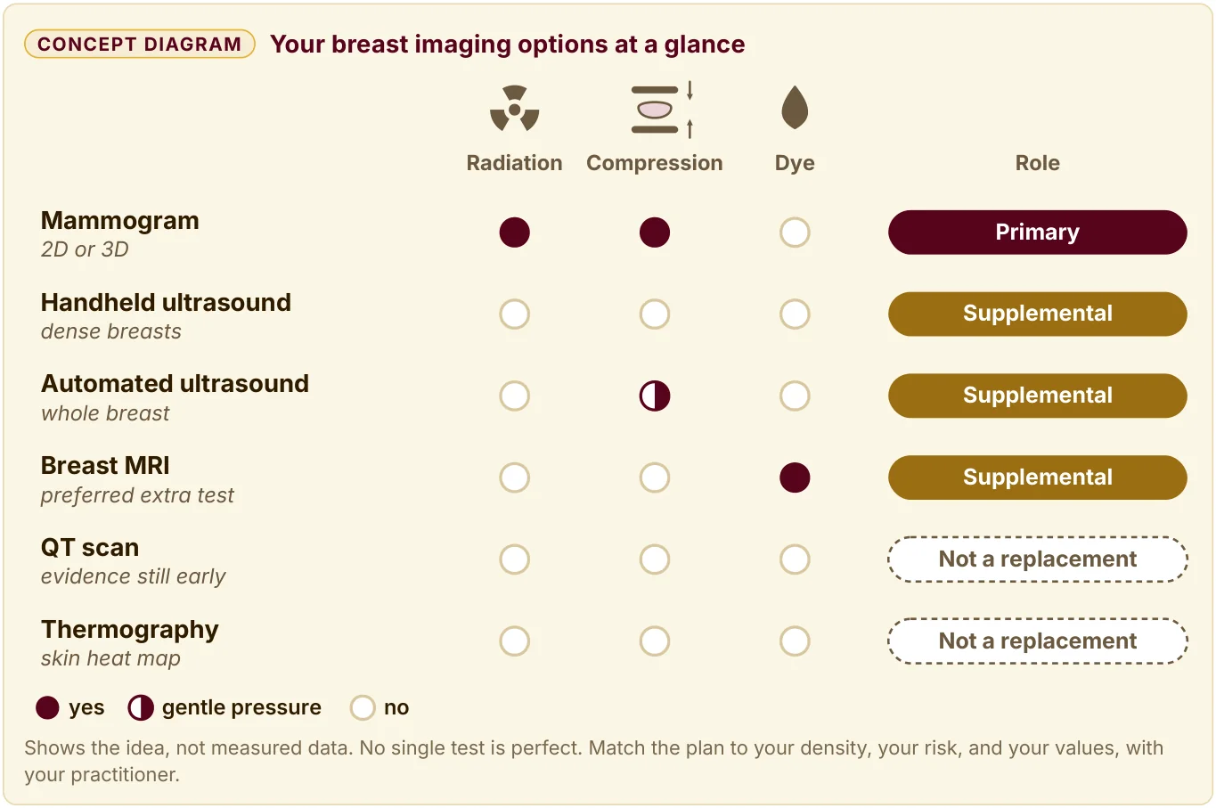
A concept diagram titled 'Your breast imaging options at a glance', with columns for radiation, compression, dye, and role. Mammogram, 2D or 3D: radiation yes, compression yes, dye no; role: Primary. Handheld ultrasound, dense breasts: radiation no, compression no, dye no; role: Supplemental. Automated ultrasound, whole breast: radiation no, gentle pressure, dye no; role: Supplemental. Breast MRI, preferred extra test: radiation no, compression no, dye yes; role: Supplemental. QT scan, evidence still early: radiation no, compression no, dye no; role: Not a replacement. Thermography, skin heat map: radiation no, compression no, dye no; role: Not a replacement. Key: a filled circle means yes, a half circle means gentle pressure, an open circle means no. Shows the idea, not measured data. No single test is perfect. Match the plan to your density, your risk, and your values, with your practitioner.
Handheld breast ultrasound. A technologist or radiologist moves a small probe over the breast, using sound waves to see inside. There is no radiation, no compression, and no contrast dye.
In dense breasts, ultrasound can find cancers a mammogram misses. In the ACRIN 6666 trial, published in JAMA in 2012, Women with dense breasts and elevated risk had yearly mammography and ultrasound. Adding ultrasound found 3.7 more cancers per 1,000 screens in the second and third years, and raised the share of cancers caught from 52 percent with mammography alone to 76 percent with both.
It also brought more false alarms. Handheld ultrasound depends heavily on the skill of the person holding the probe, so where you go matters.
Automated whole-breast ultrasound (ABUS). A wider transducer sweeps across the breast in a standard way and records a 3D volume that a radiologist reviews afterward.
In 2012, the FDA approved the first automated breast ultrasound system for use together with mammography in Women with dense breasts. In the SomoInsight study of 15,318 Women with dense breasts, adding ABUS to mammography found 1.9 more cancers per 1,000 Women, most of them invasive, along with a rise in recalls.
Breast MRI. MRI uses a magnet and radio waves, with no radiation, and it usually needs an IV contrast agent called gadolinium. MRI is the most sensitive breast imaging test in routine use, and the ACR calls it the supplemental screening method of choice for most Women at higher-than-average risk.
The strongest trial for dense breasts is the DENSE trial from the Netherlands, published in the New England Journal of Medicine in 2019. It followed 40,373 Women aged 50 to 75 with extremely dense breasts and a normal mammogram:
The trial measured interval cancers, so it cannot yet say how many lives supplemental MRI saves. It showed clearly that MRI finds cancers in dense breasts that mammography misses.
Abbreviated MRI. A shorter MRI protocol, sometimes called 'fast MRI,' shortens the scanning time compared with a full exam, and it may cost less.
In a 2020 JAMA study of 1,444 Women with dense breasts, abbreviated MRI found invasive cancer at 11.8 per 1,000 Women, compared with 4.8 per 1,000 for 3D mammography. Abbreviated MRI caught 95.7 percent of cancers, compared with 39.1 percent for 3D mammography, with more false alarms. Ask your imaging center whether it offers it and what it costs out of pocket.
What the guidelines say about supplemental imaging. The ACR's 2023 recommendations say that for Women with dense breasts who want supplemental screening, breast MRI is recommended, and contrast-enhanced mammography or ultrasound could be considered for those who cannot have MRI. The USPSTF, in 2024, concluded that the evidence is insufficient to weigh the benefits and harms of supplemental ultrasound or MRI for Women with dense breasts.
The American Cancer Society notes that experts do not agree on which extra tests, if any, Women with dense breasts should have.
So the experts disagree, and your letter says other tests may help. That combination is exactly why this conversation belongs in your hands.
Before any machine, there are your own hands and eyes.
ACOG, the American College of Obstetricians and Gynecologists, recommends breast self-awareness: knowing the normal look and feel of your own breasts, and telling your practitioner about any change. It does not recommend a formal, scheduled breast self-exam for average-risk Women, because studies did not show a benefit and it led to more false alarms.
Breast self-awareness means knowing your normal well enough to notice when something changes. Many Women notice a change in the shower, while dressing, or during a castor oil pack.
Changes to bring to your practitioner promptly:
Any new change deserves imaging, whatever your usual screening plan is.
A great treatment for your breasts, and a simple 1, is a castor oil pack.
The How It All Connects chapter mentions castor oil packs as gentle support for the liver. I use them on my breasts, too, 1 or 2 times a week.
How I do it:
Practical care:
Between my breast ultrasounds and my QT scans, castor oil packs are how I tend my own breasts at home. That is my experience. Please hold my story alongside the science.
There is no published research showing that castor oil dissolves breast calcifications. Calcifications are common, most are benign, and a small number are an early sign of DCIS or cancer.
Any new calcification needs imaging follow-up first, as your radiologist advises, before you try anything at home. Castor oil packs can be part of how you care for your breasts. They do not replace a diagnostic workup.
This article is a section of The Midlife Bible: The Essential Guide to the Women's Midlife Journey, by Daniela Hess, MSEd. It is the book's own text, so where it says 'this book' or points to another part, that means the Bible. The book has the full chapters, charts, and the Lab Wish List.
Educational content, not medical advice. Please work with your own practitioner before you change a medication or a supplement.
These 3 connect to what you just read.
Hormone therapy is 1 of the most effective tools for the Midlife years and 1 of the most misunderstood. Here is what happened in 2002, what changed in 2025, and what the evidence shows on timing, bones, heart, and breast cancer.
Read this article →Your hormones and your thyroidPerimenopause can begin as early as 35 and run up to 17 years. Here are the 3 stages, how a trained practitioner reads where you are, and why 5 hormones never work alone.
Read this article →Your heart, bones, brain, and breastsHeart disease takes roughly 7 times as many Women's lives as breast cancer, and Menopause changes your cholesterol, your blood vessels, and your body fat. Here is how a heart attack can look in a Woman, which tests to ask about, and what protects your heart every day.
Read this article →FAQ
Dense breasts have relatively more glandular and fibrous tissue and relatively less fat. You cannot feel density, and only a radiologist looking at a mammogram can tell you. Categories C and D count as dense, and in a study of about 1.5 million mammograms, 43.3 percent of US Women aged 40 to 74 had heterogeneously or extremely dense breasts.
Since September 10, 2024, every mammography facility in the United States must tell you in writing whether your tissue is dense. The letter says other imaging tests in addition to a mammogram may help find cancers. It does not schedule the next test or say which test.
Yes to both, and the second matters more for most Women. On a mammogram, fat looks dark, and dense tissue and cancer both look white. In Women 40 to 49, the sensitivity of mammography fell from 81.2 percent with non-dense breasts to 63.3 percent with dense breasts.
Compared with a Woman of average density, heterogeneously dense breasts carry about 1.2 times the lifetime risk, and extremely dense breasts about 2.1 times. The National Cancer Institute notes that Women with dense breasts who are diagnosed do not have a higher risk of dying from breast cancer than Women with fatty breasts, once other factors are taken into account.
The experts disagree, and your letter says other tests may help. The ACR's 2023 recommendations say breast MRI is recommended for Women with dense breasts who want supplemental screening. The USPSTF, in 2024, concluded the evidence is insufficient to weigh the benefits and harms of supplemental ultrasound or MRI.
In the DENSE trial of 40,373 Women aged 50 to 75 with extremely dense breasts and a normal mammogram, MRI found 16.5 additional cancers per 1,000 Women screened, along with 79.8 false-positive results per 1,000 screenings. The trial measured interval cancers, so it cannot yet say how many lives supplemental MRI saves. Mammography every 2 years for Women aged 40 to 74 remains the USPSTF screening recommendation. Every screening decision is yours to make with your practitioner.
Ask which category you are in, and which extra imaging fits your density, your risk, and your values. Ask whether your imaging center offers abbreviated MRI and what it costs out of pocket. A strong family history, a genetic mutation, or a past biopsy showing atypia makes that conversation matter even more.
If you started hormone therapy since your last mammogram, your category may change, so let the imaging center know. Most prescribers want current breast imaging on file, so if you choose a path without mammograms, talk with your prescriber ahead of time about which imaging they will accept. Hormone Therapy 101 covers the research.
Bring these to your practitioner promptly: a new lump or thickening in the breast or armpit, skin dimpling, puckering, redness, or skin like an orange peel, a change in the size or shape of 1 breast, a rash or scaling on the nipple, a nipple that newly turns inward, nipple discharge, especially if bloody or from 1 side only, and a new area of pain that stays in 1 spot. Any new change deserves imaging, whatever your usual screening plan is.
Castor oil packs can be part of how you care for your breasts. They do not replace a diagnostic workup, and any new calcification needs imaging follow-up first.

A 75-page PDF guide plus the 1 hour 43 minute talk, The Entire Midlife Journey.
Send me the guideYour thyroid, your gut, your hormones, and your labs.
Get The Midlife Bible3 hours with Daniela Hess and Kim Beekman, recorded live. The Midlife Bible is included.
See the workshop60 to 90 minutes one-on-one. Send a request to work with Daniela.
Work with Daniela